The Imaging core of the Stroke Institute provides neuroimaging services for all areas of stroke and cerebrovascular disease. Our core is divided into an acute imaging core led by Dr. Sunil Sheth and a stroke recovery section led by Dr. Muhammad Haque.
Acute Stroke Neuroimaging Research
Dr. Sunil Sheth
现在,比以往更多,神经影像让我们to look inside the brains of our patients to diagnose disease before it begins, or limit its progression once it does. Under the direction of Dr. Sunil A. Sheth, MD the UTHealth Institute for Stroke and Cerebrovascular Disease Acute Imaging Core Lab is at the forefront of this progress. Housed in a state-of-the-art facility, the Core Lab serves as a resource to local, national, and international academic collaborators studying stroke. The Lab also works with multiple industry partners to help develop the next generation of therapies for neurological disorders. Driven to fulfill its mission of “Personalizing Brain Health Through Imaging,” the Imaging Core also leads a paradigm-shifting research program using machine learning coupled with clinical data and imaging, to improve the quality of care that patients with brain injury receive regardless of whether they are treated at a small community hospital, or large academic center.
You can contact Dr. Sheth via email atsunil.a.sheth@uth.tmc.edu.
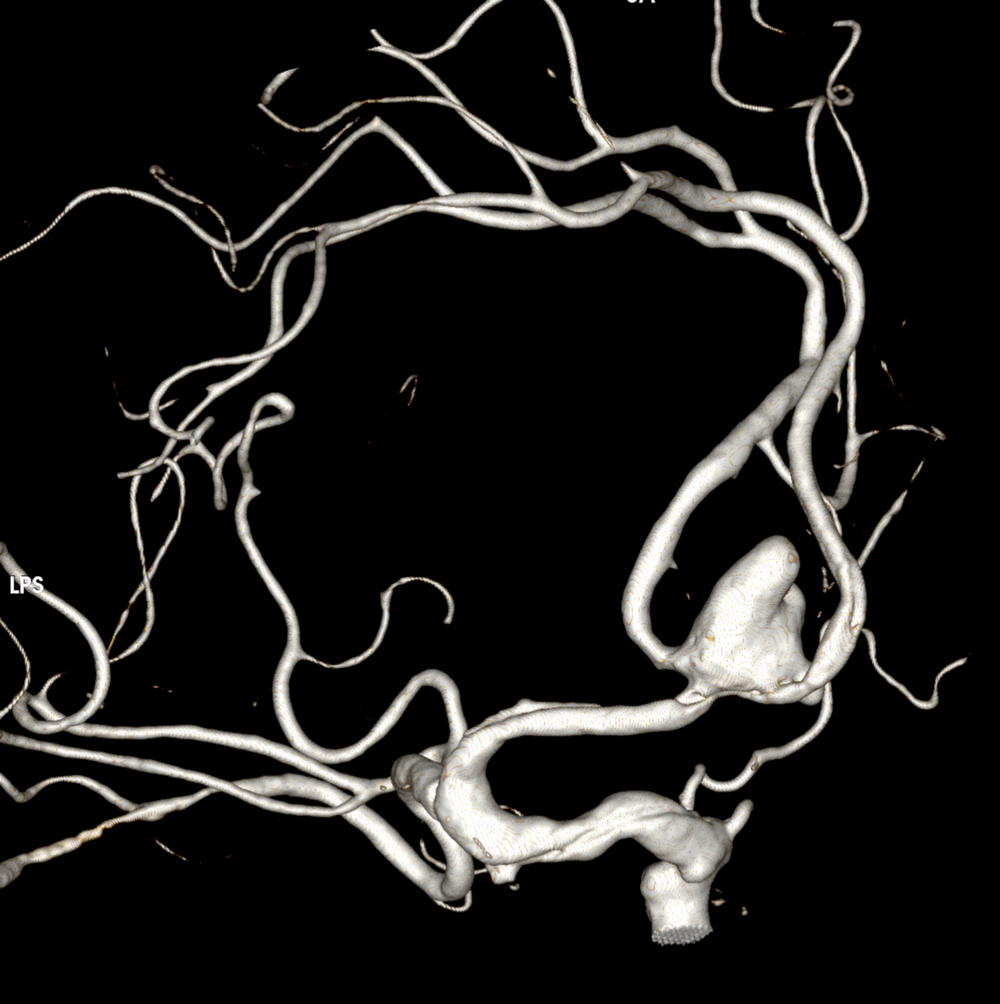
Aneurysm
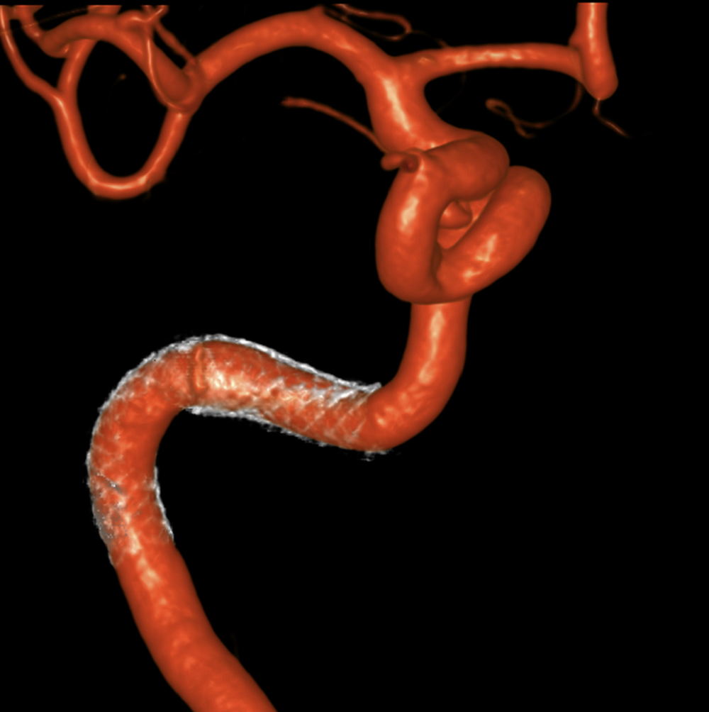
Aneurysm
Clinical and Translational MR Stroke Imaging Core
Dr. Muhammad Haque
The primary emphasis of the clinical and translational MR stroke imaging core is to develop qualitative and quantitative surrogate markers of the post-stroke prognosis, secondary degeneration, recovery, and treatment efficacy.The neuroimaging scientists at the Magnetic Resonance Image Processing Stroke Core are ready to assist you with your clinical and preclinical imaging needs by designing experiments, optimizing image acquisition protocol, and data analysis. Imaging protocols are designed to evaluate ischemic and hemorrhagic post-stroke lesion volume, white matter integrity, chronic neuronal loss, rate of brain atrophy, cerebral blood perfusion, neuronal circuitry, and metabolic profile.
Typical image acquisition protocol includes: high resolution 3D anatomical images, diffusion tensor imaging, arterial spin labeling, quantitative susceptibility imaging, magnetic resonance spectroscopy, resting state functional MRI.
Quantitative image processing includes: Voxel based morphometry, diffusion tensor matrix, deterministic and probabilistic tractography, cerebral blood volume and cerebral blood flow, lesion segmentation, neuronal network, cortical thickness, iron concentration and metabolite concentration.
我们运用基于matlab的公关ocessing code using commercial and freeware software packages such as Analyze, e-film, SPM, FSL, DTI_Studio, STI etc.
The Imaging Core also serves as the imaging analysis center for international stroke trials including the following:
- A Double-Blind, Controlled Phase 2b Study of the Safety and Efficacy of Modified Stem Cells (SB623) in Patients With Chronic Motor Deficit From Ischemic Stroke – Sunovion and San Bio, Inc
- A Double-Blind, Controlled Phase 2 Study of the Safety and Efficacy of Modified Stem Cells (SB623) in Patients With Chronic Motor Deficit From Traumatic Brain Injury (TBI) – San Bio, Inc
- Sedation versus General Anesthesia for Endovascular Therapy in Acute Ischemic Stroke – a Randomized Comparative Effectiveness Trial (SEGA)
Brain Hemorrhage and Recovery
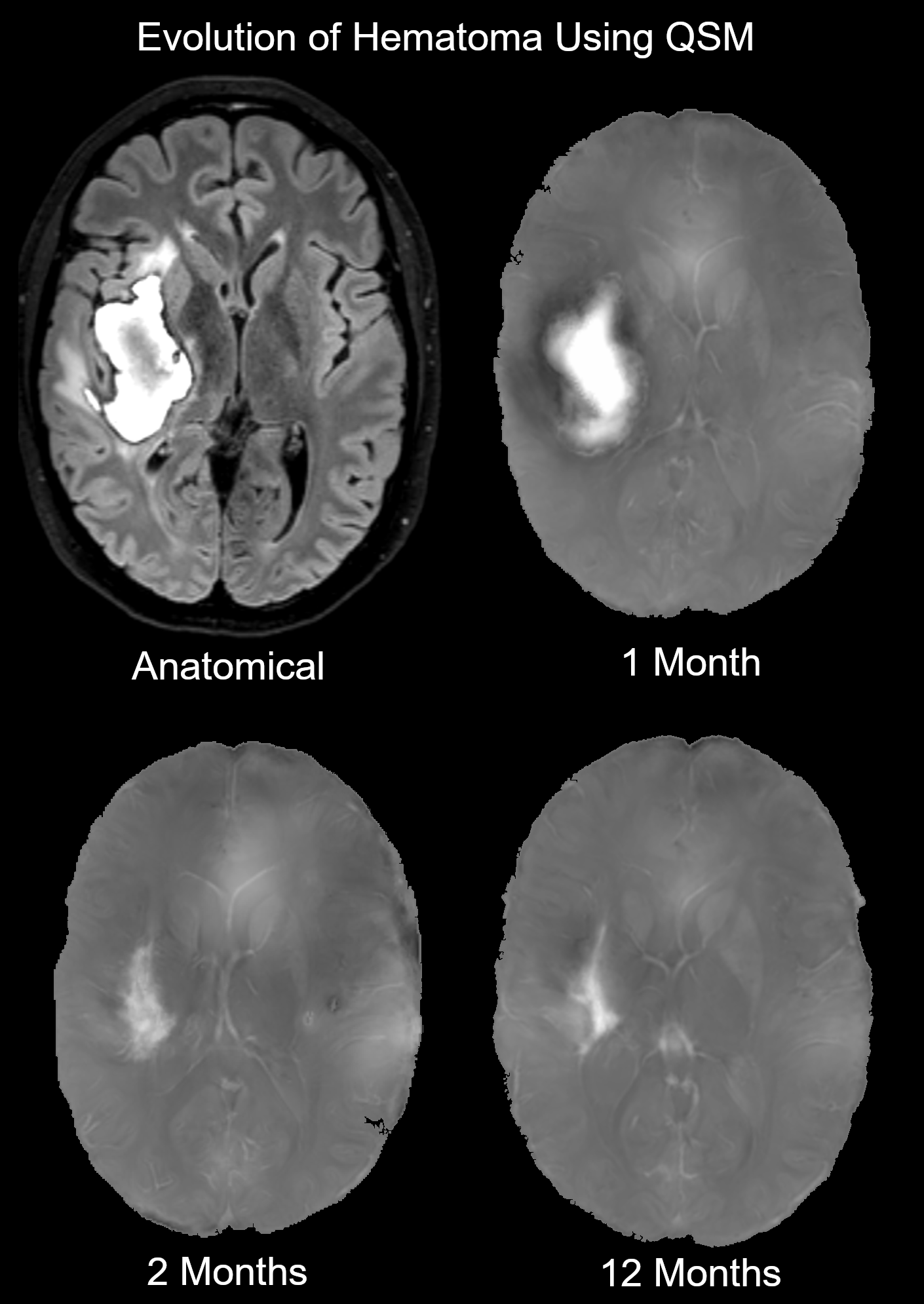
Quantitative Susceptibility Mapping allows for quantitative volumetrics of hematoma in patients with intracerebral hemorrhage
Ischemic Stroke and Recovery
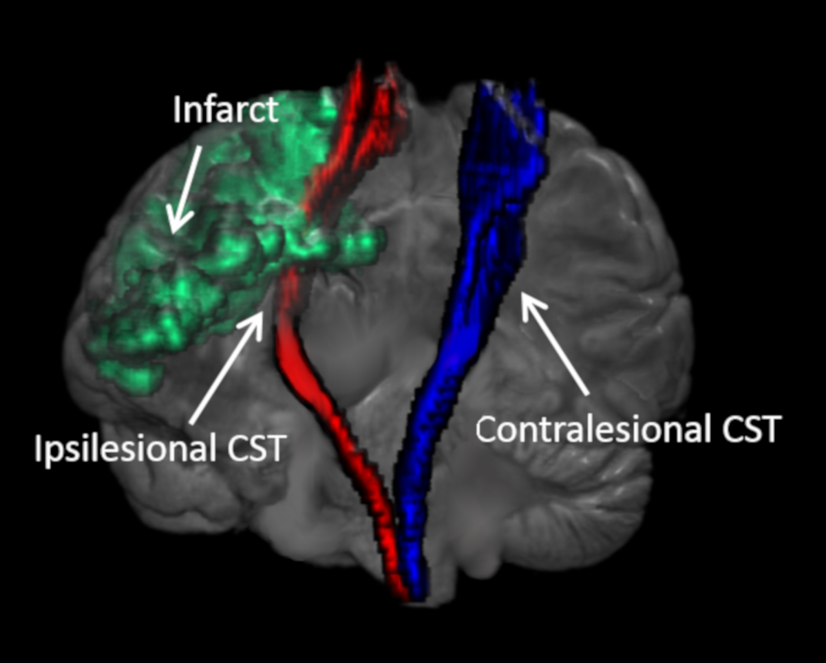
Tractography of the corticospinal tracts
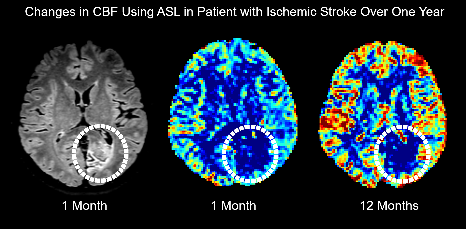
Picture of cerebral blood flow
ICH Imaging
Video of hemorrhagic stroke obtained in acute phase. Yellow, semi-translucent object represents the edema volume. Red object represents the hematoma volume. Blue object represents the ipsilesional and contralesional Corticospinal Tracts (CST).
Imaging Team
Ponnada A. Narayana, PhD, MSc
Director
Professor, Diagnostic and Interventional Imaging
Muhammad Haque, PhD
Assistant Professor, Department of Neurology
Sunil Sheth, MD
Assistant Professor, Department of Neurology
Luca Giancardo, PhD
Assistant Professor, School of Biomedical Informatics
Clark W. Sitton, MD
Associate Professor, Diagnostic and Interventional Imaging